
(3) Compared with other optical methods that have been applied to characterize breast calcifications, such as up-conversion raster scanning microscopy and laser-induced fluorescence spectroscopy, (4−6) Raman scattering is unique in revealing the intrinsic chemical compositions of tissues and biominerals.

Raman spectroscopy, on the other hand, offers a superb power of chemical identification based on the fingerprint spectral features of molecular bond vibrations. Hence, developing a method of combining the chemical and histological features of breast calcifications is crucial to the improvement of diagnostic accuracy.

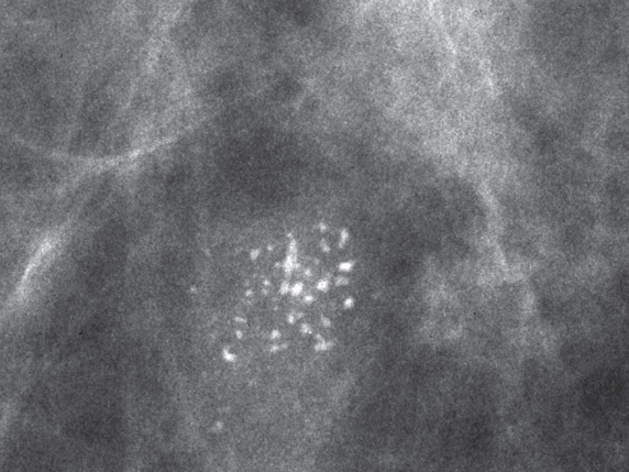
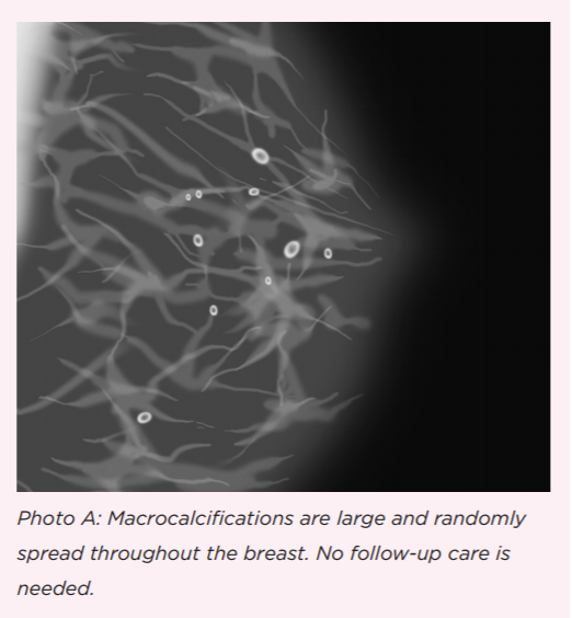
However, X-ray mammography only provides the morphological features of calcifications without any chemical resolution, which is limited for accurate tumor evaluations. (2) These calcium deposits contain rich mixtures of chemical compositions (such as phosphates, oxalates, and carbonates), as well as various morphologies (size, shape, and distributions) that are closely related to tumor development and malignancy. X-ray mammography is currently the major screening tool for early detection, (1) in which calcification is considered as a significant feature in the disease assessment and interpretation. (1) Early and accurate diagnosis of tumor malignancy could alleviate many challenges for reducing the morbidity and mortality as well as the financial burden related to the disease. Our findings may provide a rapid means to accurately evaluate breast tumor malignancy based on fresh tissue biopsies.īreast cancer is the second leading cause of cancer-related death in women and accounts for ∼23% of all cancer cases annually. With optimized combinations of chemical and geometrical features of microcalcifications, we were able to reach a precision of 98.21% and recall of 100.00% for classifying benign and malignant cases, significantly improved from the pure spectroscopy or imaging based methods. A total of 211 calcification sites from 23 patients were imaged with SRS, and the results were analyzed with a support vector machine (SVM) based classification algorithm. Here, we applied hyperspectral stimulated Raman scattering (SRS) microscopy to extract both the chemical and morphological features of the microcalcifications, based on the spectral and spatial domain analysis. In contrast, spontaneous Raman spectroscopy offers detailed chemical analysis but lacks the spatial profiles. Traditional X-ray mammography provides the overall morphologies of the calcifications but lacks intrinsic chemical information. Precise evaluation of breast tumor malignancy based on tissue calcifications has important practical value in the disease diagnosis, as well as the understanding of tumor development.


 0 kommentar(er)
